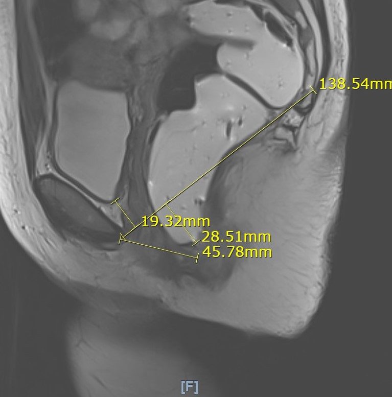
MRI (Magnetic Resonance Imaging) Proctography is a functional imaging test of the defecation process. It is ordered by your surgeon or proctologist but performed at the radiology department. It is a dynamic scan, which means not only pictures are produced but video of bodily movement.
This video test gives very valuable information about the causes of difficulties at defecation, like poor coordination or poor relaxation of the anal muscles and pelvic floor; rectocele, rectal intussusception, rectal prolapse and pelvic floor descent. It also shows how the urinary bladder and the uterus moves during straining, allowing for diagnosing cystocele and other types of POP (pelvic organ prolapse).
What to expect when going for this procedure?
At the start of the test the rectum needs to be filled up with a gel contrast through a small tube and an adult diaper is placed on. The test starts with 10-15 minutes of anatomical scans, while you are staying still, followed by a few minutes of dynamic scans, during which you are requested to tighten (squeeze) your bottom muscles then to evacuate the rectum (empty the jelly in the diaper).
No special preparation is needed for the test, but it is good to have some urine in the bladder, however, should not have an uncomfortably full bladder. To achieve this, the best is to drink as you would normally do, to stay hydrated, and go to pee 1.5-2 hours before the procedure. This allows just enough urine to build up in the bladder. The procedure is not painful, no sedation needed.
The result of the scan is visible straight after the scan, but measurements needs to be done on the images, therefore the final report is only available 3 – 7 days after the scan is done. It is advised you set up a free follow-up with your doctor to discuss the images and the plan of treatment after you have done the test.
